What is EDS?
The full name of EDS is energy dispersive X-ray spectrometer, which can record all X-ray spectra at the same time to measure the functional relationship between X-ray intensity and X-ray energy.
It is a rapid method of micro area composition analysis without damaging the sample.
Qualitative analysis of elements is carried out by measuring the characteristic X-ray energy excited by the material, and quantitative analysis is carried out by measuring the characteristic X-ray intensity.
EDS began to be commercialized in the early 1970s, and now it is basically the standard equipment of SEM.
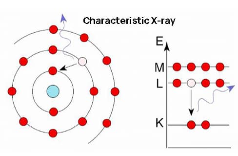
Characteristic X-ray
Definition: electromagnetic radiation photons with characteristic energy generated by the transition from the outer shell electrons to the inner shell electrons after the inner shell electrons of the atom are ionized.
When the high-speed moving electron beam bombards the sample surface, the electrons collide with the atomic nucleus and outer layer electrons of the element for one or more elastic and inelastic collisions.
About 1% of the incident electron energy excites various signals from the sample that reflect the sample information: secondary electrons, characteristic X-rays, continuous X-rays, auger electrons, backscattered electrons, etc.
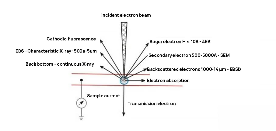
Fig. 1: signal generated by high-energy electron bombardment on sample surface
Structure and working principle of EDS
The characteristic X-ray is special because the X-ray energy released by different elements is different, just like the fingerprint of the same person, with uniqueness.
The elemental analysis using different characteristic X-ray energies is called energy dispersion method.
The structural schematic diagram is as follows:
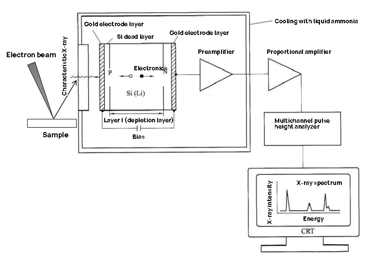
Fig. 2: structural schematic diagram of energy spectrometer
The characteristic X-ray excited by the sample is directly irradiated on the Si (LI) semiconductor detector through the window, ionizing the Si atom and generating a large number of electron hole pairs, the number of which is proportional to the X-ray energy, namely:
N = E / ε,
Wherein, ε-the energy (3.8 eV) generated to generate an electron hole pair.
For example: FeKα- the energy is 6.403keV and 1685 electron hole pairs can be generated.
By biasing the Si (LI) detector (generally – 500 ~ – 1000 V), the electron and hole pairs can be separated and collected, converted into current pulses by the preamplifier, then converted into voltage pulses by the main amplifier, and then sent to the multi-channel pulse height analyzer.
The output pulse height is determined by N, forming the abscissa of the EDS spectrum: energy.
According to the number of characteristic X-rays recorded in different intensity ranges, the intensities of X-rays of different elements can be determined to form the ordinate of EDS spectrum: intensity.
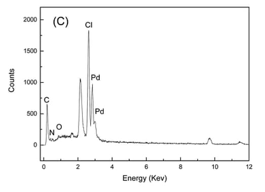
Fig. 3 EDS diagram
Range of EDS analysis elements
The elements that can be analyzed by the energy spectrometer are affected by the type of window material.
The traditional beryllium window can only analyze the elements after sodium (Na) because it absorbs the X-ray of ultra light elements.
The organic film ultra-thin window can analyze all elements between (Be) – uranium (U).
Reliability
Some people always think that EDS is a semi quantitative analysis, and the result deviation will be large.
In fact, the actual EDS is the most convenient, fast, accurate and reliable analysis method for micro region composition analysis, and the stability and reproducibility of the data are good.
Its accuracy is second only to WDS, which can reach 2-10%.
The quantitative error of the main element with no overlapping peak of the median atomic number is 2-3%, and the detection limit is 0.1-0.5%.
Generally, the reliability decreases as the atomic number decreases and the element content decreases.
The measurement depth is in the micron level.
The progress of silicon drift detector (SDD), large solid angle detector and various software processing also further reduces the measurement error of EDs.
Sample requirements
The energy spectrometer has no special requirements on the surface of the sample.
The dry solid and the stage can be placed without magnetism, radioactivity and corrosion.
If the conductivity of the sample is poor, it can be sprayed with gold or carbon.